Left atrial appendage (LAA) closure LAA image 1
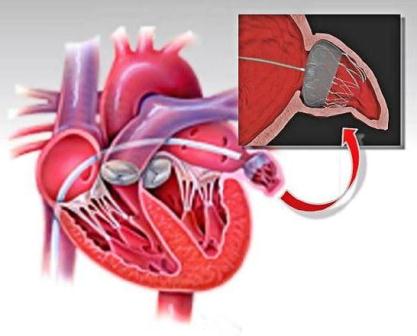
This is a treatment to prevent stroke caused by blood clots forming in the left atrial appendage in patients who suffer from atrial fibrillation (AF) and other factors that increase the risk of stroke, particularly if they have difficulty managing blood thinning therapy.
 
Patients who have artificial heart valves or who are likely to need valve replacement may not be suitable.
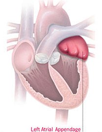
LAA image 2

The left atrial appendage is a small, windsock-like pouch, which empties into the left atrium of the heart.

For patients with normal heart rhythm, the left atrial appendage squeezes rhythmically with the rest of the left atrium. This rhythmic contraction ensures that blood in the pouch is ejected when the left atrium empties into the left ventricle, where it is then pumped all over the body.

In atrial fibrillation that rhythmic contraction is lost and blood can collect in the left atrial appendage and form a clot.

For older patients, atrial fibrillation (AF) is a common rhythm disturbance where the top chambers of the heart (atria) do not beat regularly.

The symptoms of AF vary from mild fatigue to dizziness to chest pain or difficulty breathing. Some patients feel their heart palpitating, while others are unaware of the change in heart rate.  However, whether AF causes symptoms or not the blood inside the pouch (appendage) is not ejected as it is during normal rhythm and blood clots may form in the pouch as a consequence.

These clots can pass into the left atrium and then travel to the brain causing a stroke. It is estimated that the clots which cause stroke in patients with AF originate in the left atrial appendage 90% of the time.

Most patients who develop AF are required to take "blood thinners" such as warfarin for the rest of their lives to prevent blood clots forming. Warfarin can be a difficult medication to take as there are many interactions with food items, alcohol and other drugs.

The medication dose may need frequent review and blood testing is required at least monthly. There are also significant bleeding risks while taking warfarin, particularly as patients get older. Newer "blood thinners" which are easier to take are becoming available. The bleeding risks with the newer agents are less than with warfarin but still not negligible. 
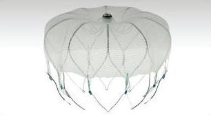
LAA image 3

An alternative approach to reduce the risk of blood clot formation in the left atrial appendage is to remove the appendage from circulation by closing it off using a form of “plug” such as the Watchman device. 
 

Before the procedure

Fasting Please stop eating six hours before your procedure time. You may continue to drink clear fluids (water, weak BLACK tea and weak BLACK coffee) up to two hours before your procedure time. Do not consume milk as this is considered food. You may take your regular medications with sips of water.

Allergies Please tell Intra staff at the time of booking if you have any food or medication allergies or previous reactions to X-ray contrast (dye).

Diabetes If you are a diabetic, please tell Intra staff at the time of booking. You may need to discuss your insulin/diabetes medication dose with your cardiologist.

Anticoagulation (blood thinner) If you are taking a blood thinner (e.g. warfarin, rivaroxaban, dabigatran, etc.), please advise Intra staff at the time of booking who will instruct you regarding continuation.

Other regular medications Continue these unless advised otherwise by your cardiologist. If you are taking a diuretic or water pills (e.g. frusemide, spironolactone) please advise Intra staff as you may need to withhold this on the morning of the procedure.

During the procedure

A breathing tube will be put in place to support ventilation and you will be asleep with for the procedure.

The femoral veins in the groin area provide the easiest venous access into the heart. This area will be prepared with an antiseptic solution. You will be covered with a large sterile drape and the area at the top of the leg will be completely numbed with local anaesthetic. This will sting for about thirty seconds.

A transoesophageal echocardiogram (TOE) probe will then be positioned in your throat operated by another cardiologist to guide the interventional cardiologist with the device positioning.

A small hollow tube called a sheath will be inserted into the vein in the groin. This will serve an as access site for the thin long flexible tube called a catheter to make its way into the heart. A needle will be used to puncture through the heart wall from the right atrium to access the left atrial appendage. The TOE probe and X-ray will be used to position accurately the occluder device. Several images with contrast dye will be taken before deployment.

Once safety checks are confirmed, the catheter and sheath are removed from the groin. Pressure is applied at the puncture site followed by a small sterile dressing.

After the procedure

Following the procedure, you will be transferred to CIU to recover and must remain lying flat on the bed for the next few hours.

Before going home you will have an echocardiogram performed to check the position of the new occluder device.

You will be required to stay in the hospital overnight, and if appropriate you will be discharged the next morning.

What are the risks?

As with any procedure, there are potential risks involved. Your cardiologist will explain the procedure, discuss possible risks and answer any questions you may have. You will then be asked to sign the consent form. This will occur either at an earlier appointment, or on the day of your procedure.