Coronary angiography
.JPG)
Coronary angiography is an examination of the coronary arteries Coronary Arteries lie on the outside of the heart and take blood to the heart muscle. These vessels are only a few millimetres wide. Just as an engine needs petrol, the heart needs blood to do its job of pumping blood around the body.
Coronary artery stenosis is the narrowing of a coronary artery. Slow build-up of fatty plaque within the artery wall can cause the artery to narrow, reducing blood flow. Sudden changes in the plaque may cause angina to worsen or may cause a heart attack.
Coronary angiography identifies the presence, extent and location of coronary artery narrowings. Other tests performed during the coronary angiogram include measuring pressures within the heart chambers, checking function of some of the valves and checking how well the main pumping chamber (left ventricle) is working.
If a narrowing suitable for stenting is found, the interventional cardiologist may then insert a stent. This procedure is referred to as Percutaneous Coronary Intervention (PCI).


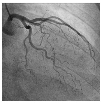
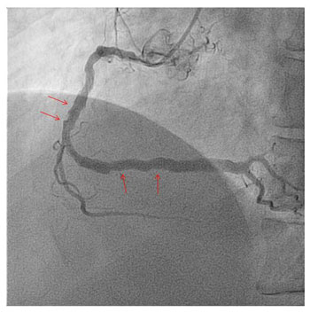
The first image shows a normal Left Coronary Artery (LCA) which supplies blood to the left side of the heart. The second image shows disease (arrows) in the Right Coronary Artery (RCA) which supplies blood to the right side of the heart.